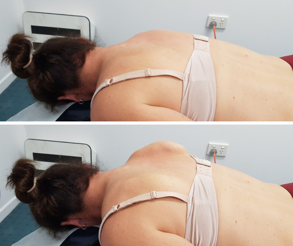
What is Neuralgic amyotrophy?
Neuralgic amyotrophy (NA) was first described by Feinberg in 1897. Patients present with sudden-onset pain in the shoulder region, followed by patchy flaccid paralysis of muscles in the shoulder and/or arm. NA is also known as “Parsonage–Turner syndrome” after the names of the researchers who first reported this disorder in detail in 1948. Many cases of NA involve the brachial plexus, and NA is thought to develop because of sudden inflammation; hence, this disorder is also known as brachial plexitis or brachial neuritis.
How common is Neuralgic amyotrophy?
In many studies, NA has been described as a rare disorder. However, recent reports suggest that NA is significantly underdiagnosed in clinical practice and has an actual incidence rate of about 1 in 1000 per year1. Although individuals of any age can be affected, NA onset usually occurs between 20 and 60 years. The paediatric incidence of NA varies according to age (from 3 days to 15 years of age) and has a biphasic peak of onset; the first is in the neonatal period, and the second peak is in adolescence (7 to 15 years).2 Patients in the acute stage of NA usually visit primary local clinics or emergency rooms, and NA is often misdiagnosed as rotator cuff tendinopathy, cervical radiculopathy, glenohumeral bursitis, or muscle strain.
What are the causes?
Although the exact pathophysiological mechanisms of NA have not yet been established, multiple factors are involved. Immunological, mechanical (e.g., repetitive strain or strenuous exercise), and genetic factors are all known to be associated with the development of NA.3
Over 50% of patients with NA have a history of an event that triggered the immune system, such as infection, vaccination, surgery, pregnancy, or physical or mental stress.
What are the symptoms?
NA often involves only one limb; however, in 10% to 30% of patients, NA is bilateral (typically in an asymmetric fashion).4 The initial symptom of NA in 96% of patients is an acute onset of pain (within a few hours) in the shoulder girdle.3 This pain usually radiates to the neck, arm, and forearm. A small proportion (1%–2%) of patients with NA have pain in a restricted area, such as the neck, scapula, or upper arm only.4 In about 60% of cases, the episodes initiate at night.3,4 Many patients therefore wake up early in the morning with severe pain, which then gains maximal intensity over the next few hours. The pain is usually of a “sharp”, “stabbing”, “throbbing”, or “aching” nature, and its intensity is typically relentless, with a numerical rating scale score ≥7 (0: no pain, 10: worst pain that a human can imagine).3,4 The pain is commonly aggravated by movement of the shoulder or limbs.3,4 The duration of pain caused by NA usually varies from several hours to several weeks before it subsides, and the average duration of pain is 4 weeks. In approximately 5% of patients, the pain resolves within 24 hours, while in 10% of patients, the pain persists for more than 2 months.3,4
Muscle weakness is a conspicuous finding in NA, and occurs days to weeks after the onset of symptoms.3,4 It characteristically worsens when the pain becomes less severe. After the onset of pain, weakness appears within 24 hours in about 30% of patients, and it occurs within 2 weeks of the pain initiation in approximately 70% of patients. In approximately 30% of patients, weakness is manifested >2 weeks after the initiation of pain. In about 70% of patients, weakness occurs in the muscles innervated by the upper trunk of the brachial plexus, either with (50%) or without (20%) involvement of the muscles innervated by the long thoracic nerve.3 The next most common location in NA is the middle and lower trunk of the brachial plexus. In addition, the lumbosacral plexus and the anterior and posterior interosseous, cranial, and phrenic nerves are peripheral nerves that are frequently involved.
Approximately 70% to 80% of patients manifest sensory deficits during episodes of NA; however, these deficits are usually mild compared with the degree of weakness.4 Hyperesthesia and/or paresthesia are the most common sensory symptoms of NA, and hypoesthesia can also occur.4 The deltoid and lateral upper arm regions are the most common sites of sensory deficits, accounting for about 50% of all such deficits. Pure sensory NA without motor impairment occasionally occurs (e.g., sural and superficial radial sensory nerves).3
How is it diagnosed?
A diagnosis of NA is based on a patient’s clinical history and physical examination.30 However, other possible disorders must be excluded. To confirm a diagnosis of NA and exclude other disorders, electrophysiological and radiographic studies are conducted. Using electrophysiological studies, the site of the lesion in the peripheral nerves can be localized, and the degree of involvement can be evaluated.31 However, during the acute stage, abnormal findings are not observed in electrophysiological studies: abnormal findings are only manifested after 1 and 3 weeks of onset of NA in nerve conduction studies and electromyography, respectively. In nerve conduction studies, there are reduced amplitudes of compound muscle action potentials in the involved nerves. Abnormal sensory nerve conduction study findings are observed in 30% to 45% of all patients with NA.32 On electromyography, there is reduced recruitment (which can be seen in acute and subacute cases), positive sharp waves, and fibrillation potential in the muscles innervated by the affected peripheral nerves.
On conventional MRI, no abnormal findings are usually apparent. However, gadolinium-enhanced MRI can be helpful for the diagnosis of NA because inflammatory sites in the involved nerves show high signal intensity.7,33 Before confirming a diagnosis of NA, MRI of the spine and ultrasound or MRI of the shoulder should be performed to rule out radiculopathy caused by a herniated disc or spinal stenosis and rotator cuff tear, respectively.13 In addition, high-resolution magnetic resonance neurography is helpful for the diagnosis of hourglass-like constriction neuropathy (a subtype of NA).8,22
Among routine laboratory investigations, the erythrocyte sedimentation rate and complete blood count are usually normal.
Treatment of Neuralgic amyotrophy
Because patients with NA experience severe pain, the active management of pain using various analgesic drugs is necessary; however, the options for complete recovery are very limited. The treatment for NA varies according to the phase in which the patient is seen. Two weeks of corticosteroid therapy is the best treatment during the painful stage—it hastens pain relief and increases the chances of recovery after 1 year.35 NA treatment usually includes a combination of corticosteroids, analgesics, immobilization, and physical therapy. Standard regimens for NA are supportive, and include a combination of analgesics (non-steroidal anti-inflammatory drugs, opioids, or neuroleptics) and immobilization to minimize pain during activities in the initial phase.36 In cases with extensive NA, intravenous corticosteroids and immunoglobulins have been used to improve functional outcomes.37 Intravenous immunoglobulin therapy with methylprednisolone pulse therapy may help to decrease symptom duration.37 If NA is diagnosed more than 1 or 2 months after onset, the pain has usually lessened, and symptomatic pharmacological treatments are usually sufficient. However, in severe cases, opioids, corticosteroids, and intravenous immunoglobulins may still be considered.38 Most cases do not require analgesics during the palsy phase; however, in cases with neuropathic pain, specific neuropathic pain relief medications may be required. In other cases, residual pain is related to muscular compensation caused by weakness within the palsied muscles, and rehabilitation is necessary. In addition, compensatory orthotic or surgical procedures may be considered in cases of persistent weakness.
Surgical options include intrafascicular neurolysis and neurorrhaphy/grafting; however, many authors recommend waiting for at least 3 months for spontaneous recovery because many patients show spontaneous recovery during this period. If no clinical signs of recovery are noted in 3 months, magnetic resonance neurography is recommended.35,39 If nerve constriction is revealed, surgery can be considered. If the percentage of nerve thinning caused by nerve constriction is less than 75%, intrafascicular neurolysis is recommended. When nerve constriction is ≥75%, neurorrhaphy/grafting should be considered.40
Prognosis for Neuralgic amyotrophy
The prognosis of NA depends on the degree of collateral reinnervation. Overall, patients with NA recover to 80% to 90% of their previous state in 2 to 3 years; however, >70% of patients with NA have residual motor weakness. Electrodiagnostic studies and evaluations of motor strength can be helpful for predicting the prognosis of patients with NA. If the amplitude of compound motor action potentials is reduced by more than 70% or there is an initial paresis of grade ≤3 on the Medical Research Council scale for muscle strength, collateral reinnervation will be incomplete and the prognosis is likely to be poor. Overall recovery was less favourable than usually assumed, with persisting pain and paresis in approximately two-thirds of the patients who were followed for 3 years or more.
Physiotherapy management
Physical therapy (PT) focuses on educating and training movement and position sense, coordination of the affected shoulder girdle and improving functional endurance. Occupational therapy (OT) focuses on prevention and reduction of overuse of affected and compensating muscles, body ergonomics at rest and during activities and adaptation of activities and environmental changes.
PT included training to regain scapular muscular balance and progressive resistance training of rotator cuff muscles; the latter only after scapular muscular balance was achieved since scapular stability is essential for the function of arm muscles that control position. All exercises were carried out without or only with minimal pain during and after exercises. If patients did experience (excessive) pain during or after the exercises, intensity of the program was adjusted accordingly. If patients had difficulty in implementing the scapular control movements in daily life, scapular proprioceptive taping was used to increase awareness of their scapular position during posture and movement. Some patients with NA experience neural stretching pains of the brachial plexus, which are likely caused by neural entrapment in case of a habitually protracted and adducted scapula because of serratus anterior weakness and compensatory activation of the pectoralis and trapezius muscles. When present, this complication was treated by increasing scapular control as described above and by using neural mobilisation techniques, using movements designed for the Upper Limb Tension Test. These involved patients to grab hold of the doorframe and stretch out their arm until they experienced mild neurological sensations such as tingling or radiating pain. They were instructed to perform this stretching. exercise three times a day in three repetitions, lasting 20 to 30 seconds. Patients who lacked control of their primary cervical stabilizing muscles, tested using the cranio-cervical flexion test with pressure biofeedback, received sensomotory cervical stability training. When increased muscle tone and/or myofacial trigger points were found in the neck or shoulder region patients were treated with muscular relaxation exercises and trigger point releases as pre-conditioning for sensomotory scapular and cervical training.
The focus of the OT intervention lied on enabling daily occupations. Patients gained insight into activities that provoked pain and into strategies focussed on preventing and reducing pain caused by overuse of affected and compensating muscles. To this end, energy conservation strategies were taught including taking mini-breaks, practicing optimal body ergonomics during rest and action, and analysing and adapting activities or the environment with or without use of aids and adaptations (Ghahari et al., 2010). Patients learned self management strategies to reduce stress and physical strain and to find a balanced distribution of activities during the day/week.
In males the initial pain tended to last longer than it did in females (45 versus 23 days). In females the middle or lower parts of the brachial plexus were involved more frequently (23.1 versus 10.5% in males), and their functional outcome was worse.
As always if you have any questions always give us a call to talk through any concerns you have regarding NA or other shoulder disorders.
References
- Van Alfen N, Van Eijk JJJ, Ennik T, et al. Incidence of neuralgic amyotrophy (Parsonage Turner syndrome) in a primary care setting – a prospective cohort study. PLoS One2015; 10: e0128361. [PMC free article] [PubMed] [Google Scholar]
- Rotondo E, Pellegrino N, Di Battista C, et al. Clinico-diagnostic features of neuralgic amyotrophy in childhood. Neurol Sci2020; 41: 1735–1740. [PubMed] [Google Scholar]
- Van Alfen N. Clinical and pathophysiological concepts of neuralgic amyotrophy. Nat Rev Neurol2011; 7: 315–322. [PubMed] [Google Scholar]
- Van Alfen N, Van Engelen BG. The clinical spectrum of neuralgic amyotrophy in 246 cases. Brain2006; 129: 438–450.


