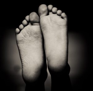
It is not usually until you have to have an x-ray that you may find you have an extra bone in your skeletal make-up of your feet. Although sometimes you may notice that your foot has a mysterious bump that other friends and family do not have.
Do not freak out!!! Breathe! It is a normal variation!
Accessory bones or Ossicles of the foot are a normal variation that can present as both symptomatic and asymptomatic. There are normally only 26 bones in each foot and an extra bone can mean the foot shape can vary from the average making it difficult to accommodate the extra bone in footwear. This can cause rubbing and therefore irritation of the extra bone or structure that it is sitting within e.g. a tendon.
Another reason pain can occur is if the bone potential gets in the way functionally. For example there is an accessory bone (Os Trigonum) that can occur at the back of the ankle joint and when the foot/ankle is maximally pointed, such as in activities such as en pointe in ballet or kicking in football, it can “get in the way”, impinge and cause pain.
Below are examples of accessory bones of the foot:
Os peroneum – occurs on the lateral part of the foot area of the cuboid bone. It sits within the peroneus longus tendon and is quite common occurring in up 26% of feet (Jones).
Os tibiale externum – also known as Os naviculare accessorium is usually a large accessory ossicle that presents on the medial side of the navicular bone of the foot. The tibialis tendon will often insert into the ossicle and in some people can cause a tendinosis due to the traction of the navicular and the ossicle. It is present in approximately 10% of the population (Jones).
Os trigonum – This accessory bone sits posterior to the talus bone and can sometimes be mistaken for a fracture. It occurs in approximately 7% of adults (Jones). The ossicle usually develops between the ages of 7 and 13 and usually fuses with the talus, otherwise continuing as an Os trigonum (Jones).
Bipartite hallux Sesamoid – is a normal variant that can occur in up 33% of hallux sesamoids (Jones). It is more common in the medial sesamoid than the lateral sesamoid and an important differential diagnoses is a stress fracture of the sesamoid bone (Jones). This can be difficult to differentiate because bipartite hallux sesamoids are more likely to fracture than complete hallux sesamoids (Jones) .
Os supranavicular – is located on the dorsal (top) aspect of the foot above the navicular bone or talonavicular joint. It is present in less than 1% of the population (Jones).
If there are any further questions in regards to accessory bones please contact me via email
To book with Aleks please contact us or simply call Burleigh Physio on 5535 5218
References
Jones, Jeremy. “Os Trigonum | Radiology Reference Article | Radiopaedia.Org”. Radiopaedia.org. N.p., 2017. Web. 22 Mar. 2017.
Gold Coast Physiotherapy and Allied Health at Burleigh Heads and Broadbeach


